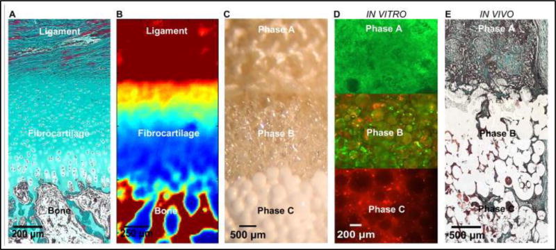
Figure 2.
a) The native ACL-bone interface exhibits distinct yet continuous tissue regions, including ligament, fibrocartilage, and bone. (Neonatal Bovine, Modified Goldner Masson Trichrome Stain, bar = 200 µm). b) Fourier Transform Infrared Spectroscopic Imaging or (FTIR-I) revealed that relative collagen content is the highest in the ligament and bone regions, decreases across the fibrocartilage interface from ligament to bone (neonatal bovine, bar=250 µm, with blue to red representing low to high collagen content, respectively). c) A tri-phasic stratified scaffold designed to mimic the three distinct yet continuous interface regions (bar=500 µm). d) In vitro co-culture of fibroblasts and osteoblasts on the tri-phasic scaffold resulted in phase-specific cell distribution and the formation of controlled matrix heterogeneity. Fibroblasts (Calcein AM, green) were localized in Phase A and osteoblasts (CM-DiI, red) in Phase C over time. Both osteoblasts and fibroblasts migrated into Phase B by day 28 (bar = 200 µm). e) In vivo evaluation of the tri-phasic scaffold tri-cultured with Fibroblasts (Phase A), Chondrocytes (Phase B), and Osteoblasts (Phase C) revealed abundant host tissue infiltration and matrix production (week 4, Modified Goldner Masson Trichrome Stain, bar = 500 µm). Adapted from (8) with permission.

