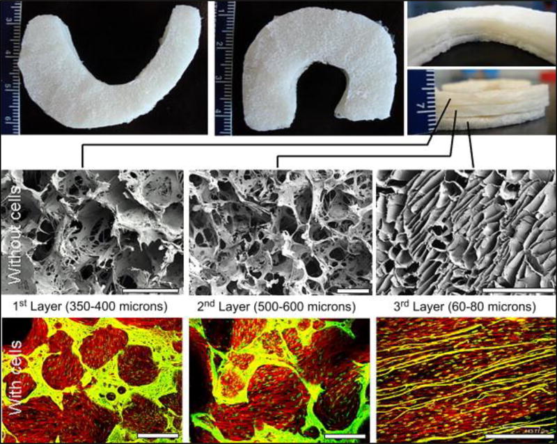
Figure 5.
Meniscus shaped scaffolds with three stacked layers (Top). SEM images showing porosity and pore interconnectivity in the individual silk layers without cells (middle) and confocal images of layers with confluent cells (bottom). Scale bar represents 500 microns. Adapted from(49) with permission.

