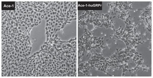
Fig. 1.
Phase contrast microscopy of Ace-1 and Ace-1-huGRPr cells showing the difference in morphology. The parent Ace-1 cells had a typical epithelial phenotype with formation of a cobblestone pattern of adherent polygonal cells. In contrast, the Ace-1-huGRPr cells had a spindle-shaped morphology and were often dissociated from one another.

