Figure 5.
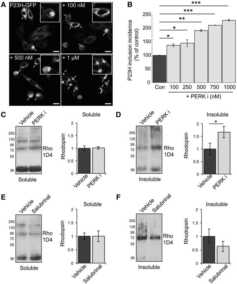
PERK inhibition increases P23H rhodopsin aggregation. (A, B) SK-N-SH cells were transfected with P23H-GFP rod opsin. Three hours post-transfection and after 2 h recovery in serum cells were either left untreated or treated with 100, 250, 500, 750 mM and 1μM of PERKi for 18 h prior to fixation. (A) Representative confocal images of P23H-GFP untreated rod opsin or treated with 100mM, 500 mM and 1 μM PERKi as indicated. Scale bar 10 μm. Magnified cell images are shown in insets. (Β) The incidence of inclusion formation of P23H-GFP in the absence and presence of PERKi at the indicated concentrations was assessed by scoring the percentage of cells with rod opsin P23H-GFP inclusions in 8 fields of ∼100 transfected cells. (C–F) Retinae of P23H-1 rats treated from P21-P35 with either 100 mg/kg PERKi (C,D), or salubrinal (E, F) or vehicle were analysed by a sedimentation assay. Fractions were immunoblotted with the 1D4 antibody against rhodopsin. Densitometric analysis was used to calculate the levels of soluble rhodopsin (C,E) relative to the vehicle treated and insoluble rhodopsin (D,F) relative to the vehicle after normalisation to soluble rhodopsin. Values are means ± SEM, n ≥ 4 (biological replicates). Error bars represent standard error, *P < 0.5, **P < 0.01, ***P < 0.001 Students t-test.

