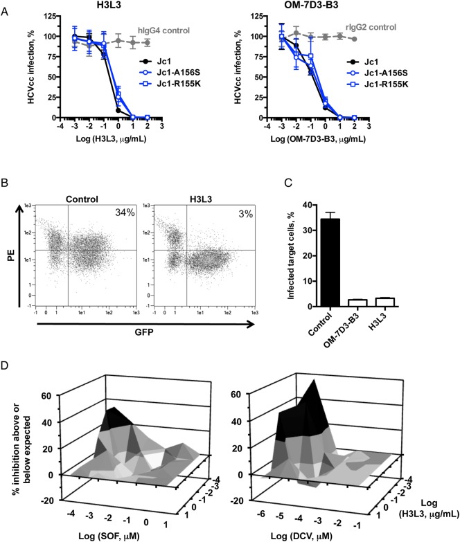
Figure 5.
Functional characterisation of the humanised anti-tight junction protein claudin-1 (CLDN1) antibody clone H3L3 in cell culture. (A) H3L3 dose-dependently inhibits HCV infection. Huh7.5.1 cells were incubated with rat OM-7D3-B3, humanised H3L3 or isotype control antibodies prior to infection with HCVcc (Jc1 wild type, Jc1-A156S or Jc1-R155K). Infection was assessed by measuring luciferase activity after 72 hours. Results are expressed as log relative luciferase units from three independent experiments performed in triplicate. (B and C) H3L3, like OM-7D3-B3, inhibits HCV cell–cell transmission. Huh7.5.1 cells electroporated with HCV Jc1 RNA (producer cells) were co-cultured with naive Huh7.5.1-GFP cells (target cells) in the presence of control or anti-CLDN1 antibody (11 µg/mL). Co-cultured cells were fixed with paraformaldehyde after 24 hours and stained with an NS5A antibody (B). The extent of cell–cell transmission was determined by calculating percentage of GFP+NS5A+ cells (C). Results from a single experiment performed in duplicate are shown. (D) H3L3 synergises with direct-acting antivirals. Huh7.5.1 cells were pretreated with H3L3 in combination with sofosbuvir (SOF) or daclatasvir (DCV) prior to infection with HCVcc. Infection was assessed after 72 hours by luciferase activity. Synergy (20% above that expected for additive effects, shown in black) was assessed according to the Prichard and Shipman method.40 Results from a representative experiment are shown.

