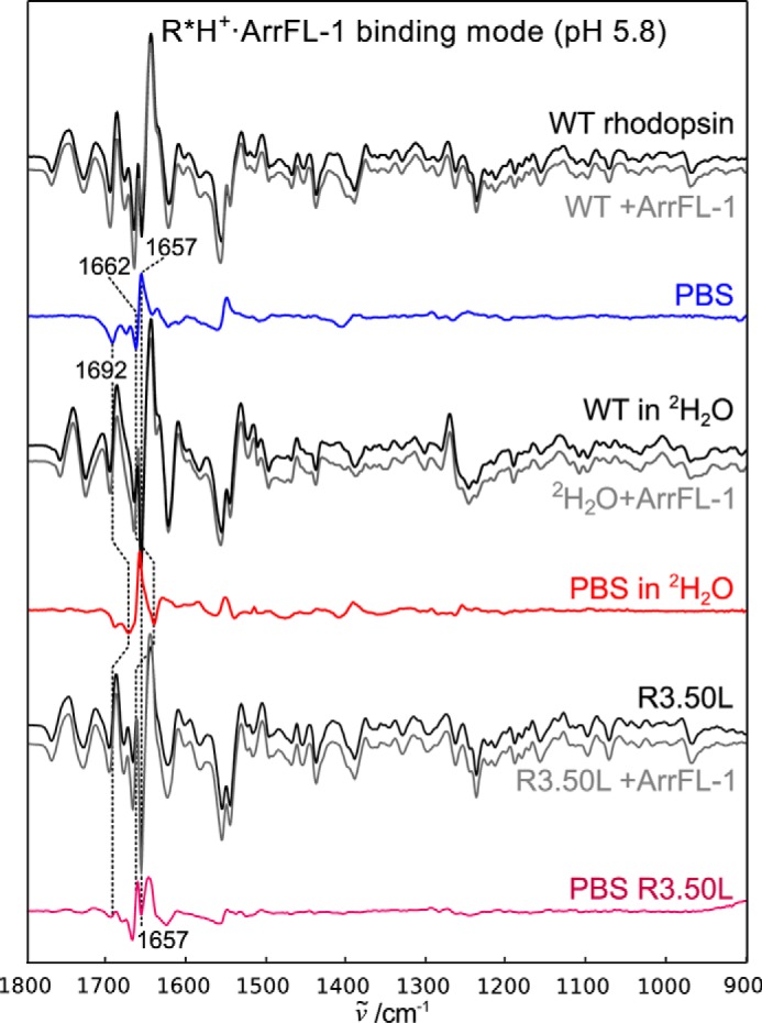
Figure 4.

The R*H+·ArrFL-1 binding mode stabilized at low pH. Difference spectra recorded in the absence of ArrFL-1 peptide are shown in black, in the presence of 20 mm ArrFL-1 in gray. The resulting PBS are shown in blue (WT), red (in 2H2O), or purple (R3.50L mutant). Note the difference in band pattern compared with the deprotonated complex. The small effect of 2H2O on the positive 1657 cm−1 indicates that this band is a structurally sensitive amide I band.
