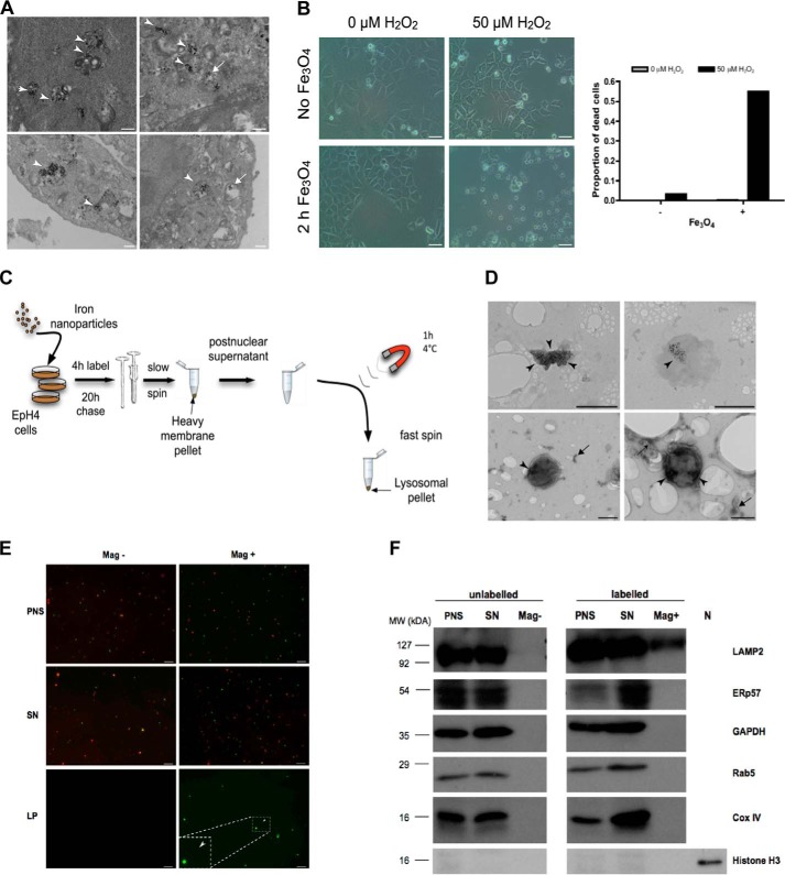
Figure 1.
Iron nanoparticle-mediated isolation of EpH4 cell lysosomes. A, TEM images of EpH4 cells showing iron nanoparticles residing in degradative lysosomal vacuoles (arrowheads) or in lysosomal vacuoles devoid of degradative material (arrows). Scale bars: 500 nm. B, bright-field microscopy and quantification of iron nanoparticle–induced cell death of EpH4 cells observed solely in the presence of hydrogen peroxide (H2O2). EpH4 cells were treated with H2O2 for 19 h. Scale bars: 25 μm. C, schematic of the magnetic iron nanoparticle lysosomal purification protocol. Adapted from Sargeant et al. (20). D, TEM images of lysosomes (top panel) and negatively stained lysosomes (bottom panel) isolated using the magnetic iron nanoparticle purification protocol. Arrowheads mark areas containing iron nanoparticles. Arrows indicate unidentifiable membranous fragments, which may be endolysosomal tubules or remnants from the ER or Golgi apparatus. Scale bars: 500 nm. E, isolated EpH4 lysosomes are predominantly clear of mitochondria. EpH4 cells were labeled with 10 kDa FITC-dextran (2.5 mg/ml (green)) with (Mag+) or without (Mag−) iron nanoparticle–containing media for 2 h and subsequently stained with MitoTrackerTM (500 nm (red)) for 30 min prior to isolation. Representative images of the PNS, post-magnetic SN, and magnetic lysosomal pellet (LP) of the two conditions are shown. Scale bar: 10 μm. F, isolated EpH4 lysosomes are highly pure with undetectable contamination from other organelles as observed by immunoblotting. N, nuclear lysate. Organelle marker proteins: LAMP2, lysosomes; ERp57, endoplasmic reticulum; GAPDH, cytoplasm; Rab5, early endosomes; Cox IV, mitochondria; Histone H3, nucleus.

