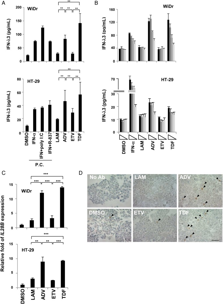
Figure 4.
Nucleotide analogues directly induce interferon (IFN)-λ3 in colon cancer cells. (A) Colon cancer cells (WiDr and HT-29) were incubated with dimethyl sulfoxide (DMSO, 0.1%), lamivudine (LAM, 50 μM), adefovir pivoxil (ADV, 2.5 μM), entecavir (ETV, 0.25 μM) or tenofovir disoproxil fumarate (TDF, 50 μM). After 48 hours, the supernatants were subjected to a chemiluminescence enzyme immunoassay for IFN-λ3. IFN-α (100 U/mL), with or without poly I:C (30 μg/mL) or R-837 (5 μg/mL), was used as a positive control. (B) WiDr and HT-29 cells were treated with each nucleos(t)ide analogue (NUC) diluted 1:3 (maximum concentration of each NUC is shown above). (C) WiDr and HT-29 cells were treated under the same conditions as above for 24 hours, and total RNA was extracted. The IL-28B mRNA expression level was determined using real-time PCR, as described in the online supplementary materials and methods. At least three independent experiments were conducted in triplicate, and representative data are shown. (D) WiDr cells were treated with 0.1% DMSO, 50 μM LAM, 2.5 μM ADV, 0.25 μM ETV or 50 μM TDF for 48 hours, and immunohistochemistry was performed. ‘No Ab’ indicates that the cells were treated without the primary antibody. Arrowheads indicate positive staining. Scale bar represents 50 μm. *p<0.05, **p<0.01 and ***p<0.001. PC, positive controls.

