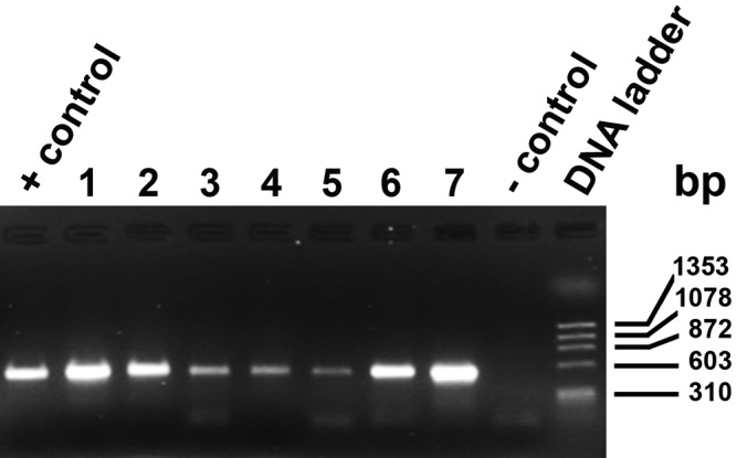
Figure 4.

Representative PCR samples separated on an agarose gel. Lane sequence: positive control (D. canis), Lane 1, whole skin (frozen); 2, whole skin (frozen); 3, whole skin (formalin-fixed); 4, whole skin (formalin-fixed); 5, whole skin (formalin-fixed); 6, tape impression, individual mite; 7, tape impression, individual mite; negative control (water), and DNA ladder. Size markers are indicated. The PCR amplicon was 537 bp.
