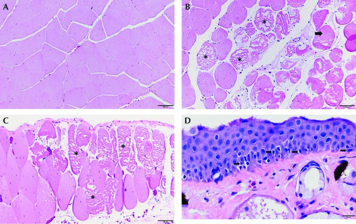
Figure 4.
Postoperative day 3. Skeletal muscle deep to the right dorsal lymph sac. (A) Normal myocytes adjacent the right dorsal lymph sac in a frog given MS222 and 25 mg/kg flunixin meglumine. (B) Degenerate and necrotic myocytes (*) in frog that received etomidate at 22.5 mg/L and 25 mg/kg flunixin meglumine. Myocytes are swollen and fragmented, with loss of cross striations. (C) Frog anesthetized with benzocaine at 0.1% and 25 mg/kg flunixin meglumine exhibits hypereosinophilic, degenerate (arrow) and necrotic, fragmented (*) myocytes. (D) Skin over the right dorsal lymph sac. Frog anesthetized with benzocaine 0.1% and 25 mg/kg flunixin meglumine. Basal keratinocytes in the skin over the dorsal lymph sac are necrotic with pyknotic nuclei (arrow). The basement membrane is intact. Hematoxylin and eosin staining; magnification, 10× (A–C); 40× (D).

