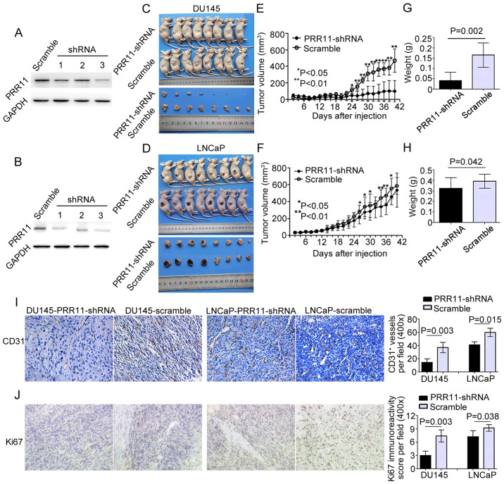
Figure 5.
Knockdown of PRR11 suppresses tumor growth and angiogenesis in vivo. (A and B) Western blotting showed that PRR11 expression in PCa cells was inhibited by three lentivirus expression plasmids containing small interfering RNAs against PRR11. The cells transfected with the third lentivirus expression plasmid were used in subsequent experiments. (C and D) Knockdown of PRR11 by the lentivirus-shPRR11 vector in DU145 and LNCaP cells inhibited subcutaneous tumor growth over the 42-day monitoring period after tumor cell injection. (E-H) The tumor growth curves are shown. Knockdown of PRR11 suppressed the growth of tumor nodules and reduced the weight of tumors in the PRR11-shRNA groups on day 42 compared with the control groups (scramble). (I and J) Immunochemistry analysis of the tumor xenografts. CD31 stained the cytomembrane or cytoplasm of pan-endothelial cells undergoing angiogenesis. Ki67 stained the nucleus of proliferative PCa cells (shown in the fields at a magnification of ×400). The CD31 staining results indicated that PRR11 downregulation reduced angiogenesis in tumor xenografts compared with control tumor xenografts. The Ki67 staining results indicated that the tumor xenografts established by cells with low expression of PRR11 expressed less Ki67 protein. The results are presented as means ± SD. *P<0.05, **P<0.01 compared with the negative control.

