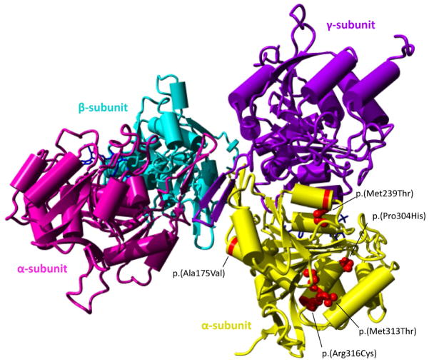
Figure 4.
3D structure of the IDH3 complex and the location of the missense variants.
The IDH3 protein complex contains two alpha subunits (pink, yellow), one beta subunit (purple) and one gamma subunit (blue). Missense variants are indicated in red in only one of the two alpha subunits for clarity. In patients with compound heterozygous variants, the NAD-IDH complex may consist of two identical alpha subunits altered by the same variant or two different alpha subunits.

