
Figure 2. Early postnatal ablation of AgRP neurons results in reduced bone mass.
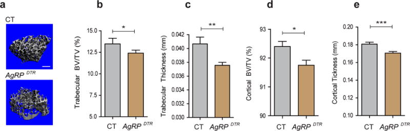
(a) Representative micro-CT images of femoral trabecular bone from three-month-old male control (CT) and AgRPDTR mice. Scale bar = 500 μm. (b–e) Micro-CT analysis demonstrated a reduction in trabecular bone volume (BV/TV), trabecular thickness, cortical BV/TV and cortical thickness in three-month-old male AgRPDTR mice (b: n=10 for CT, n=20 for AgRPDTR, p<0.05; c: n=10 for CT, n=20 for AgRPDTR, p<0.001; d: n=10 for CT, n=20 for AgRPDTR, p<0.05; e: n=10 for CT, n=20 for AgRPDTR, p<0.001). * p<0.05, ** p<0.001 and *** p<0.001. Data are presented as means ± s.e.m. P values for unpaired comparisons were analyzed by Student’s t test.