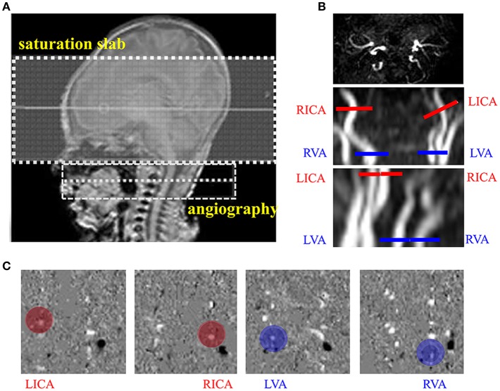
Figure 1.
Measurement of cerebral blood flow (CBF) using phase contrast (PC) MRI. (A) Positioning of magnetic resonance angiography (MRA). (B) Positioning of PC MRI for the left internal carotid arteries (LICA), right internal carotid arteries (RICA), leftvertebral arteries (LVA) and right vertebral arteries (RVA) using the maximal-intensity-projection (MIP) images of the TOF angiogram. (C) Example velocity maps of the LICA, RICA, LVA, and RVA. Blood flow velocity was measured in the velocity map (red and blue circle).

