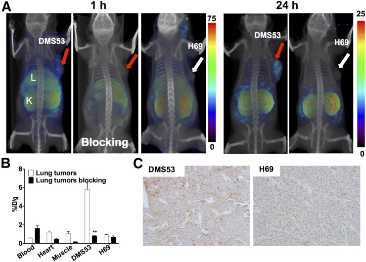
FIGURE 4.
PSMA imaging in subcutaneous lung cancer xenografts with known PSMA-specific radiotracer, 125I-DCIBzL. (A) Male NOD/SCID mice bearing DMS53 or H69 xenografts were administered 37 MBq (1 mCi) of 125I-DCIBzL via tail vein injection, and SPECT/CT images were acquired 1 h and 24 h (right panels) afterward. Arrows = tumor; L = liver; K = kidney. (B) Mice harboring DMS53 or H69 xenografts were administered 74 kBq (20 μCi) of 125I-DCIBzL, and biodistribution studies were performed 1 h afterward. For blocking dose, DCIBzL at 50 mg/kg was coinjected with 125I-DCIBzL. Data are mean ± SEM of 4 animals. Significance of value is indicated by asterisks, and comparative reference is blocking dose uptake in same tumor. **P < 0.01. (C) Representative microscopy images of PSMA-stained sections from same cohort of mice obtained at ×20 magnification.

