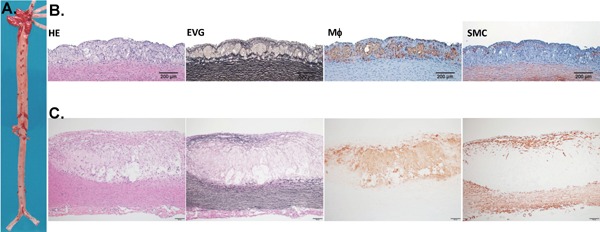
Fig. 1.

Representative aortic lesions in cholesterol-fed rabbits. A. Gross lesions of the aorta stained by Sudan IV (visualized as red area). B. Representative micrographs of early stage lesions (fatty streaks). C. A typical fibrous plaque with a lipid core covered by a fibrous cap. Serial paraffin sections of the aortic arch were stained with hematoxylin and eosin (HE) and elastica van Gieson (EVG), or immunohistochemically stained with monoclonal antibodies (mAbs) against either macrophages (M?) or α-smooth muscle actin for smooth muscle cells (SMC).
