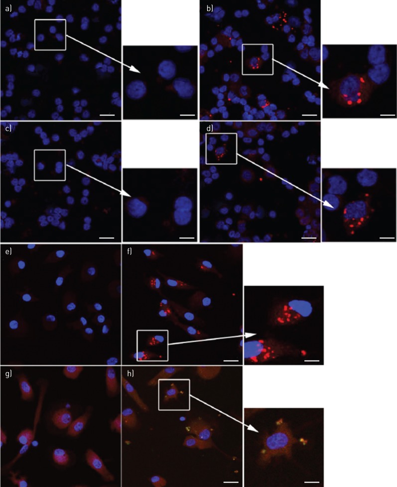
FIGURE 7.
Nontypeable Haemophilus influenzae (NTHi)-induced inflammasome specks in peripheral blood mononuclear cell (PBMC) and blood monocytes. Specks of NLRP3 (red) were not detected in a) PBMCs cultured in control medium, but were readily detected in b) the presence of NTHi. Specks of AIM2 (red) were not detected in PBMCs c) cultured in control medium, but were readily detected in d) the presence of NTHi. Specks of NLRP3 (red) were e) not detected in monocytes cultured in control medium, but f) were readily detected in the presence of NTHi. g) and h) Dual labelling of NTHi-stimulated monocytes revealed colocalised specks of AIM2 (red) and cleaved interleukin (IL)-1β (green). Yellow is merged colour of red and green. Blue is the pseudocolour of DAPI (4',6-diamidino-2-phenylindole). Scale bars=20 μm (main images) and 8 μm (inset images). Images are representative confocal microphotos of cells from of at least four donors.

