
Figure 6.
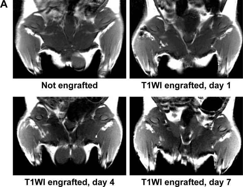

(A) T1WIs of rats before MNP-Mn(II)-labeled BMSCs were injected into the right hip. T1WIs of rats after MNP-Mn(II)-labeled BMSCs were injected into the right hip on days 1, 4, 7, 14, 21, and 28. Hyperintense T1 signals in the right hip represented the presence of engrafted MNP-Mn(II)-labeled BMSCs in vivo. (B) The relative signal intensity of the MNP-Mn(II)-labeled BMSC injection region and the adjacent soft tissue over time. (C) The area of time-dependent hyperintense signals generated by MNP-Mn(II)-labeled BMSCs.
Abbreviations: BMSCs, bone marrow-derived stem cells; MNP-Mn(II), manganese (II) ions chelated to melanin nanoparticles; MRI, magnetic resonance imaging; PBS, phosphate-buffered saline; WI, weighted image.