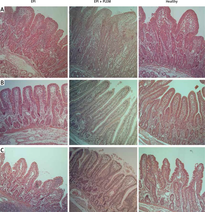
Figure 2.
Microphotographs of hematoxylin-eosin stained mucosal samples from proximal (A), middle (B) and distal (C) parts of small intestine
EPI group – pigs with pancreatic duct ligation (n = 4), EPI + PLEM group – pigs with pancreatic duct ligation and dietary supplementation with PLEM (pancreatic like-enzymes of microbial origin) for 7 days (n = 5), Healthy group – pigs with intact pancreas (n = 6). Magnification 400×. Bar 100 μm.

