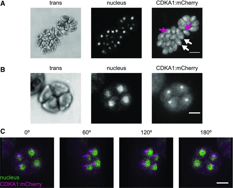
Figure 6.
CDKA1 Is Localized to the Nucleus and the Base of Flagella.
Confocal micrographs from a population of live, cycling cdka1ΔCDKA1:mCherry cells. Contrast was adjusted separately for each image, so intensities should not be compared across images. Cytoplasmic background fluorescence in the mCherry channel was also observed in cells lacking the transgene (data not shown).
(A) CDKA1 localization in newborn cells. CDKA1 was found in nuclei (magenta arrows) and in small puncta at the apical end of the cell body (white arrows), possibly at the base of the flagella. Images are average intensity z-projections. Bar =10 μm.
(B) In dividing cells, CDKA1 was primarily found in small puncta. Bar = 5 μm. Images are average intensity z-projections.
(C) CDKA1 puncta in dividing cells were adjacent to nuclei. Images are rotations from a brightest point 3D projection of the cell cluster in (B). Bar = 5 μm.

