Figure 5.
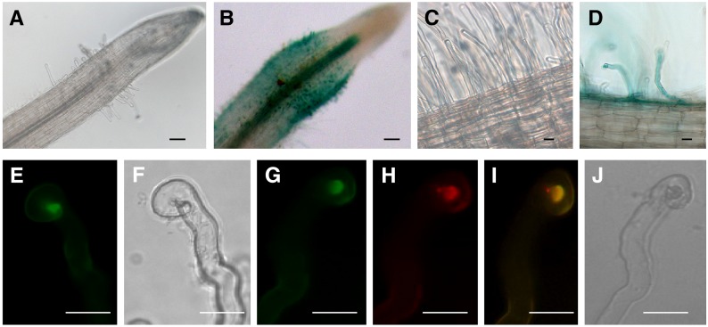
Expression of MtNFH1 in Response to Rhizobial Inoculation and Accumulation of MtNFH1:GFP in the Infection Chamber.
(A) to (D) Analysis of M. truncatula R108 roots transformed with a MtNFH1pro-GUS construct. Plants were inoculated with S. meliloti Rm41 (3 dpi; [B] and [D]) or left noninoculated ([A] and [C]). Roots were stained with X-Gluc and then cleared with diluted NaClO solution. Bars = 50 µm in (A) and (B) and 20 µm in (C) and (D).
(E) to (J) Analysis of curled root hairs of R108 roots expressing MtNFH1:GFP driven by the MtNFH1 promoter (3 dpi) under green fluorescence ([E] and [G]), red fluorescence (H), and bright-field conditions ([F] and [J]). Roots were inoculated with S. meliloti Rm41 ([E] and [F]) or S. meliloti 1021 (pQDN03) constitutively expressing mCherry ([G] to [J]). GFP fluorescence signals reflecting the presence of MtNFH1:GFP protein are increased in the infection chamber. Colocalization with bacteria is indicated in yellow (merged image; [I]). Bars = 20 µm.

