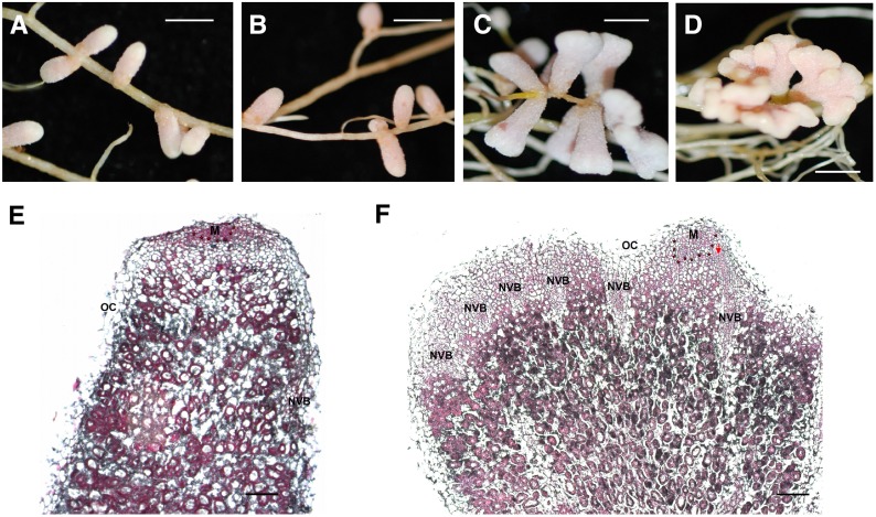
Figure 6.
Nodulation Phenotype of the nfh1-3 Mutant.
Plants were inoculated with S. meliloti Rm41.
(A) Unbranched nodules formed on M. truncatula R108 wild-type roots (20 dpi).
(B) Wild-type nodules formed on roots of a wild-type sibling line of nfh1-3 (20 dpi).
(C) and (D) Bifurcate (C) or palmate-coralloid (D) nodules formed on roots of the nfh1-3 mutant (20 dpi).
(E) and (F) Microscopy analysis of a wild-type (E) and nfh1-3 (F) nodule (20 dpi). Sections were stained with ruthenium red. M, meristem (indicated with asterisks); NVB, nodule vascular bundle; OC, outer cortex; PVM, provascular meristem (red arrow).
Bars = 2 mm in (A) to (D) and 200 µm in (E) and (F).

