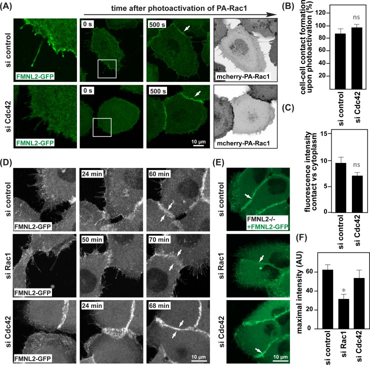
Fig 5. Rac1 is required for FMNL2 localization to cell-cell contacts.
(A) Live-cell imaging of FMNL2-/- cells expressing FMNL2-GFP together with mCherry-PA-Rac1 transfected with either control siRNA or siRNA directed against Cdc42. Red arrows highlight FMNL2-GFP localization in filopodia (B) Quantification of cell-cell contact formation as in (A). (n = 53 (si control), n = 43 (si Cdc42), pooled from three independent experiments, ns non-significant, calculated by t-test). (C) Quantification of fluorescence intensity of FMNL2-GFP at cell-cell contacts versus cytoplasm (n = 13 (si control), n = 13 (si Cdc42), pooled from three independent experiments, ns non-significant, determined by t-test.) (D) Time lapse images of FMNL2-/- cells expressing FMNL2-GFP transfected with control, Rac1 or Cdc42 siRNA. (E) Stills of live FMNL2 -/- cells expressing FMNL2-GFP transfected with control, Rac1 or Cdc42 siRNA. Arrows highlight FMNL2-GFP at cell-cell contacts. (F) Quantification of fluorescence intensity at cell-cell contacts in cells treated with siRNA as indicated (n = 8 (si control), n = 12 (si Rac1), n = 6 (si Cdc42), pooled from three independent experiments,*p≤0.01, error bars SEM). siRNA efficiency in MCF10A cells is demonstrated in S1J Fig.

