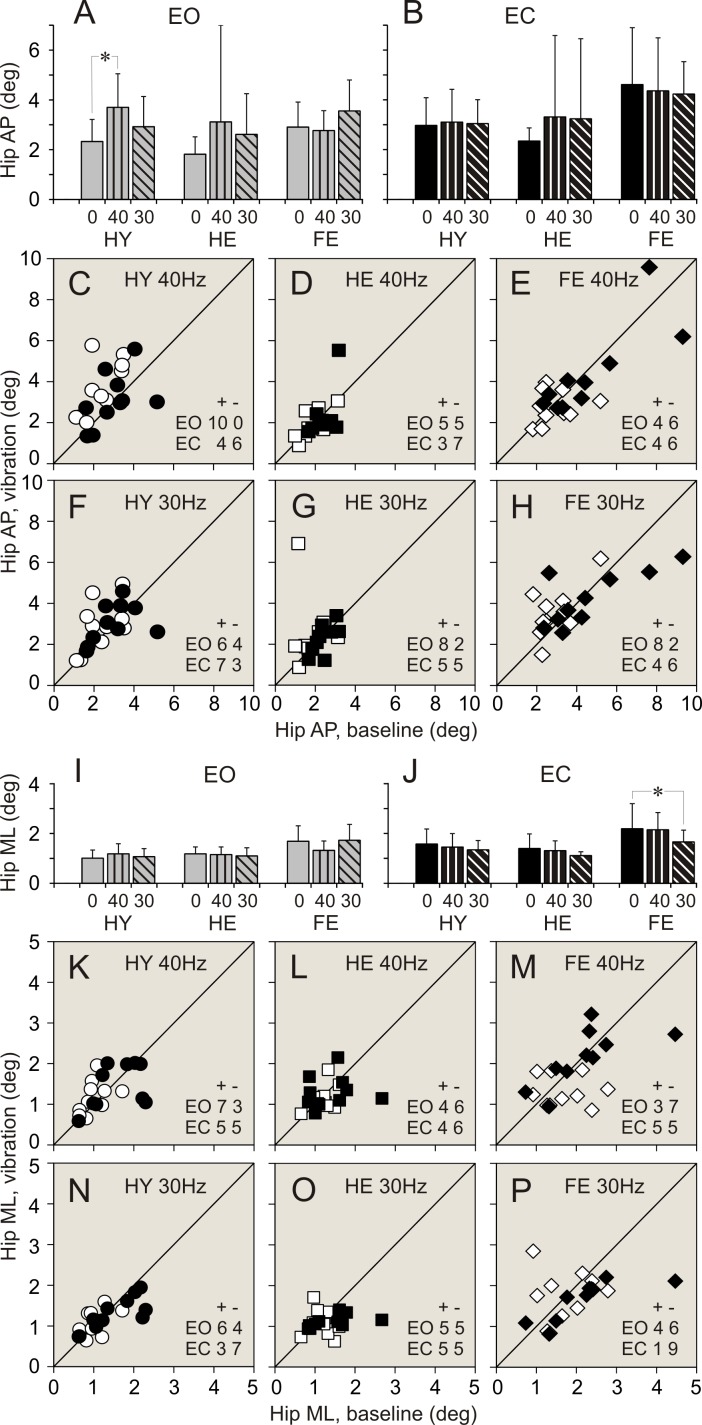
Fig 5. Hip angular deviations during standing with and without vibration of ankle muscles.
(A, B) Hip anterio-posterior (AP) deviations during standing with eyes open (EO), and eyes closed (EC), respectively. (C, D, E) Individual AP deviations during standing with 40 Hz vibration plotted against deviations during quiet standing in participants of HY, HE, and FR groups, respectively. (F, G, H) Individual AP deviations during standing with 30 Hz vibration plotted against deviations during quiet standing in participants of HY, HE, and FR groups, respectively. (I, J) Hip medio-lateral (ML) deviations during standing with eyes open (EO), and eyes closed (EC), respectively. (K, L, M) Individual ML deviations during standing with 40 Hz vibration plotted against deviations during quiet standing in participants of HY, HE, and FR groups, respectively. (N, O, P) Individual ML deviations measured during standing with 30 Hz vibration plotted against deviations during quiet standing in participants of HY, HE, and FR groups, respectively. Designations as in Fig 3.

