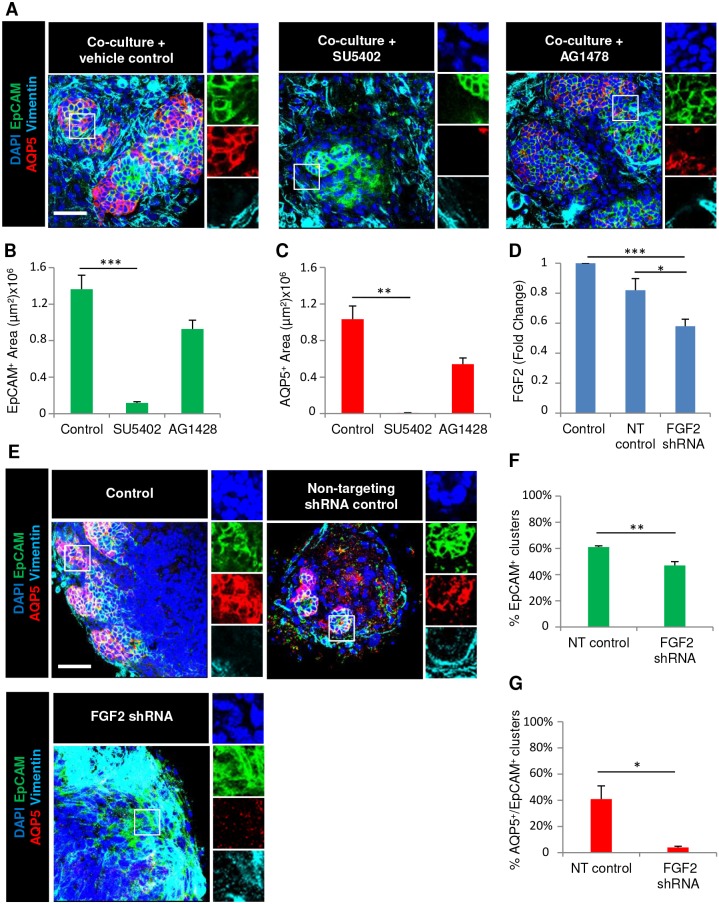
Fig. 3.
Mesenchyme-dependent salivary organoid formation requires FGF2 expression by the mesenchyme. (A) E16 epithelial clusters were seeded on primary mesenchyme feeder layers with the fibroblast growth factor receptor (FGFR) or epidermal growth factor (EGFR) inhibitors SU5402 and AG1478, respectively, in simple medium and were examined by ICC and confocal imaging. Quantitative analysis revealed loss of (B) epithelial EpCAM and (C) proacinar AQP5 protein expression with FGFR inhibition but not EGFR inhibition. (D) E16 mesenchyme cells were infected with lentiviral constructs expressing non-targeting (NT) shRNA or FGF2-targeting shRNA. An ELISA using cell lysates demonstrated a decline in FGF2 protein levels in mesenchymal cells infected with shRNA. (E) E16 epithelium was co-cultured with mesenchyme following lentiviral treatment with NT or FGF2 shRNA for 7 days. ICC and confocal imaging revealed (F) a decline in the epithelial population and (G) loss of proacinar differentiation, as shown by staining for the markers EpCAM (epithelium, green), AQP5 (proacinar/acinar cells, red) and vimentin (mesenchyme, cyan) with DAPI (nuclei, blue). *P<0.05, **P<0.01, ***P<0.001 (one-way ANOVA with Tukey post-hoc test between each condition for B–D; Student's t-test for F,G). n=3 experiments. Scale bars: 50 μm.

