Fig. 3.
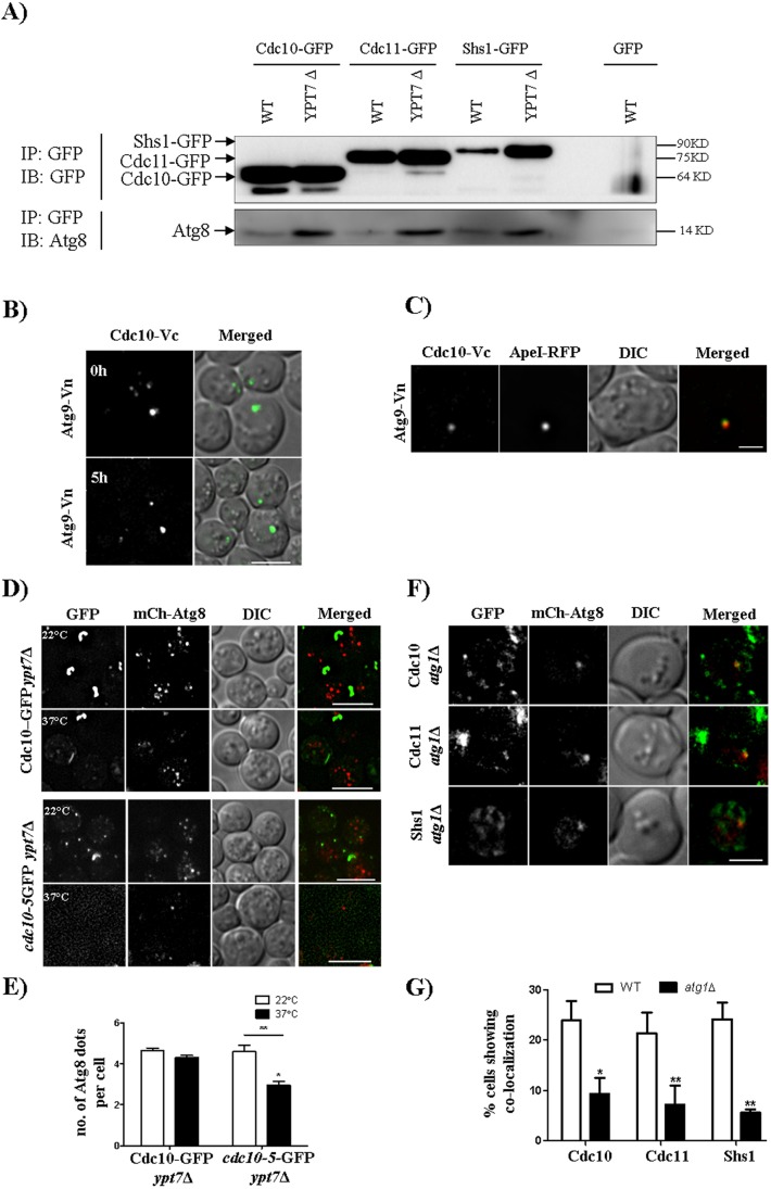
Septins are involved in autophagosomes biogenesis. (A) Western blot showing the septin–Atg8 interaction in WT and in ypt7Δ strains expressing Cdc10–GFP, Cdc11–GFP and Shs1–GFP. Cells were grown as described in the Materials and Methods. IB, immunoblot; IP, immunoprecipitation. (B,C) BiFC experiments. A strain expressing Cdc10-Vc (Cdc10 C-terminus tagged with C-terminus of Venus) and Atg9-Vn (Atg9 C-terminus tagged with N-terminus of Venus) with or without Ape1–RFP was grown as described in Fig. 1B and imaged after 5 h of incubation in starvation medium. (D) Representative images and (E) quantification showing autophagosome number per cell at 37°C. All the images are maximum intensity projections, and more than 50 cells were quantified manually with Fiji. *P<0.05 (comparison between non-Ts and Ts at 37°C); **P<0.01 (comparison between 22°C and 37°C in Ts) (two-way ANOVA). (F) Representative images and (G) quantification of colocalization events between mCherry–Atg8 and the three GFP-tagged septins in the atg1Δ strain. A total of 50 cells were quantified manually at every z-plane. *P<0.05 for Cdc10–GFP, **P<0.01 for Cdc11–GFP and Shs1–GFP (two-way ANOVA). Scale bars: 2 µm (C,F); 5 µm (B,D).

