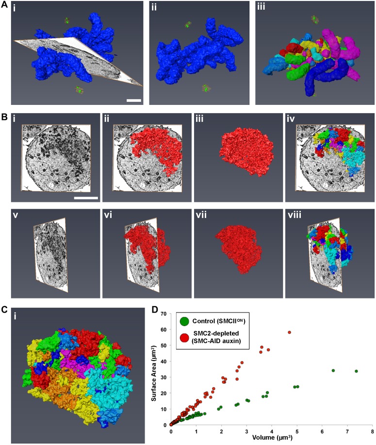
Fig. 5.
3D-EM shows that condensin is required for chromosome organization, but not for chromatin compaction. Control (SMC2ON) and condensin-depleted (SMC-AID+auxin) cells were imaged by SBF-SEM. EM data were used to generate digital models in Amira. (A) Representative images of a control cell with all chromosomes (indigo) modeled. Model is shown with (i) and without (ii) the EM orthoslice. Centrioles (green) and pericentriolar material (orange) are also shown. Individual chromosome units were identified (iii) and separated (multi-colored). (B) Representative images of a SMC2-depleted cell with all chromosomes modeled (red), shown at different angles, with and without the EM orthoslice (i–iii,v–vii). Individual chromosome units were identified and separated. Images show chromosomes traversing an orthoslice (iv and viii). (C) Enlargement of the model from Biv. (D) 2D scatter plot of surface area versus volume for all separated chromosome units of control (green) and SMC2-depleted (red) cells. Scale bars: 2 µm (A), 4 µm (B).

