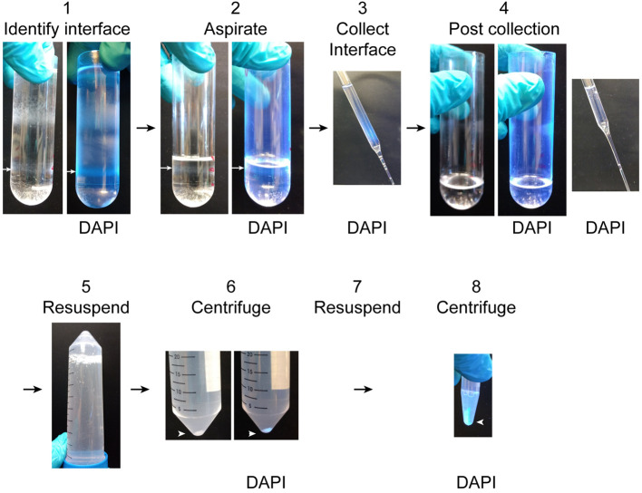
Figure 4. Unloading ultracentrifuge tubes.
1. Identify the interface between the 2.8 M and 2.1 M sucrose solutions which contains the nuclear fraction (white arrow). It is easy to identify by DAPI fluorescence under UV light. 2. Aspirate the overlying layers to a centimeter above the nuclear fraction (white arrow). 3. Collect the nuclear fraction into the BSA-coated 50 ml conical tube. Nuclei in the fraction can be observed by DAPI fluorescence. 4. After collecting the nuclear fraction, the thin gray band of nuclei will no longer be visible and no DAPI fluorescence will be detectable in the ultracentrifuge tube or sucrose collected from the surface of the remaining solution. 5. Resuspend the collected nuclear fraction 1:10 in resuspension buffer by inverting the BSA-coated 50 ml conical tube several times. 6. Pellet the purified nuclei by centrifugation. The nuclear pellet is easy to see both with and without DAPI labeling. 7. Remove the supernatant and resuspend the pellet in 1.5 ml resuspension buffer. 8. Pellet the nuclei by centrifugation. Nuclei can be resuspended for imaging or washed 2 more times for biochemical and proteomic analyses.

