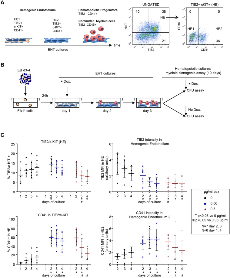
Fig. 2.
High Runx1 induction in iRunx1ko induces accelerated EHT. (A) Left: schematic of hemogenic endothelium differentiation in mESC-derived EHT cultures. HE1 (TIE2+/c-KIT+/CD41−) cells transition via the HE2 stage (TIE2+/c-KIT+/CD41+) into TIE2−/CD41+ hematopoietic progenitors and TIE2−/CD45+ committed myeloid cells. Right: flow cytometry of HE1 and HE2 populations in day 2 wild-type mESC-derived EHT cultures. (B) Schematic of hematopoietic differentiation experiments. FLK1+ cells, isolated from mESCs differentiated as EBs, were induced with doxycycline 1 day after re-plating in EHT-culture conditions. Day 2 and 3 EHT cultures were subjected to hematopoietic CFU assays with or without additional doxycycline. (C) Flow cytometry data of a 4-day time course depicting the transition through HE1 and HE2 in iRunx1ko cells after induction by low (0.06 µg/ml) and high (0.3 µg/ml) levels of doxycycline. Top left: percentage HE cells. Top right: median fluorescence intensity of TIE2+ cells in HE. Bottom left: percentage of HE2 cells within HE. Bottom right: median fluorescence intensity of CD41+ cells within HE2. Individual biological experiments and mean±s.d. are shown. Two-way ANOVA. N=biological replicates.

