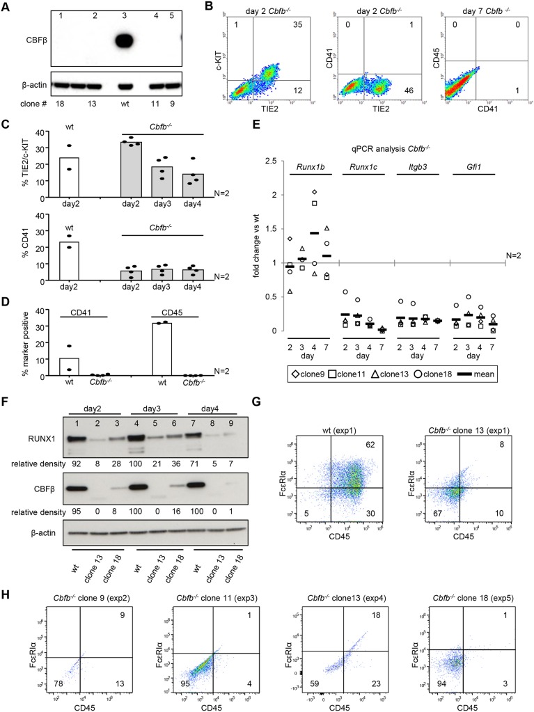
Fig. 4.
Cbfb−/− have reduced RUNX1 protein levels and cannot initiate EHT. (A) Western blot for CBFβ on CRISPR/Cas9-generated Cbfb−/− mESC clones. Lane 3 shows a wild-type (wt) mESC positive control. (B) Representative flow cytometry plots for Cbfb−/− day 2 and day 7 EHT cultures. (C-E) Four Cbfb−/− clones were differentiated across two independent experiments. Individual clones and mean are shown. (C,D) Flow cytometry data for four Cbfb−/− clones analyzed on day 2-4 (C) and day 7 (D) of EHT culture. (E) Time-course qPCR analysis of Cbfb−/− EHT cultures. Data are normalized to the wild-type line. (F) Time-course western blot analysis of EHT cultures for RUNX1, CBFβ and β-actin on two Cbfb−/− clones (lanes 2, 5, 8, and 3, 6, 9) and wild type (lanes 1, 4, 7). β-actin-normalized relative densities for RUNX1 and CBFβ are shown. (G,H) Flow cytometry analysis of four Cbfb−/− clones across five independent mast cell differentiations.

