Fig. 5.

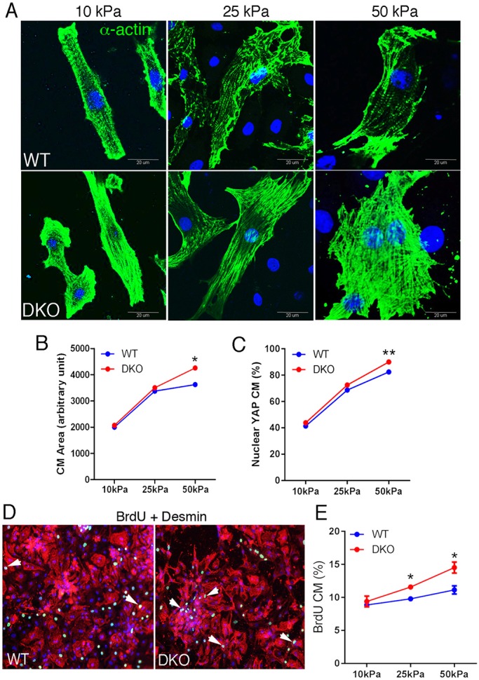
α-Catenin DKO cardiomyocytes exhibit increase proliferation on stiff ECM. (A) Representative images of neonatal cardiomyocytes cultured on different PA-FN stiffness for 4 days and stained for α-actin (green) and DAPI (blue). (B,C) Quantification of (B) cell area and (C) nuclear Yap in cardiomyocytes grown on different stiffnesses. (D) Representative images of BrdU (5-bromo-2′-deoxyuridine, green) incorporation (arrowheads) in cultured neonatal cardiomyocytes (desmin, red). Cells were co-stained for desmin (red). (E) Quantification of BrdU-positive cardiomyocytes cultured on different stiffnesses (n=3 experiments, n=minimum 100 cells/experiment). *P<0.05, **P>0.01 by Student's t-test. Error bars represent s.e.m.
