Fig. 1.
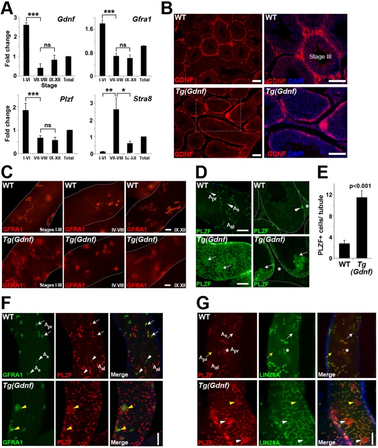
Stage-specific expression of GDNF increases the As SSC population. (A) qRT-PCR on seminiferous tubule RNA, staged by transillumination (Fig. S1A). Fold change is relative to gene expression within the total testis (arbitrary value 1). *P<0.01; **P<0.005; ***P<0.0005; ns, not significant. (B) GDNF immunostaining shows high, uniform expression in tubules of formalin-fixed sections of Tg(Gdnf) tubules. Boxed area in Tg(Gdnf) is enlarged on the right. DAPI stains nuclei. (C) GFRA1 immunostaining of whole-mount adult Tg(Gdnf) tubules shows high density of GFRA1+ cell clusters present at all stages. (D) Immunostaining in whole-mount tubules (left) or sections (right) shows large clusters of PLZF+ cells in Tg(Gdnf) mice (arrows) compared with wild type (arrowheads). Clusters are often present near interstitial spaces (asterisks). (E) Quantification of PLZF+ cells on 6-week-old testis sections (see Fig. S2A). Values represent mean PLZF+ cells±s.e.m. (n=3 per genotype). (F) In wild type, whole-mount tubule immunostaining shows co-expression of GFRA1 and PLZF in As (arrowheads) and Apr (arrows) spermatogonia. In Tg(Gdnf) testis, GFRA1+ cells cluster and co-express PLZF (yellow arrowheads). (G) Some Apr (asterisk) and all Aal chains co-express PLZF and LIN28A in wild-type whole-mount tubule immunostains. Some As (white arrows) and Apr (yellow arrows) cells do not express LIN28A. In Tg(Gdnf) tubules, the cores of PLZF-expressing clusters, show negative (yellow arrowheads) or reduced (white arrowheads) expression of LIN28A. See also Fig. S2. Scale bars: 100 µm.

