Fig. 6.

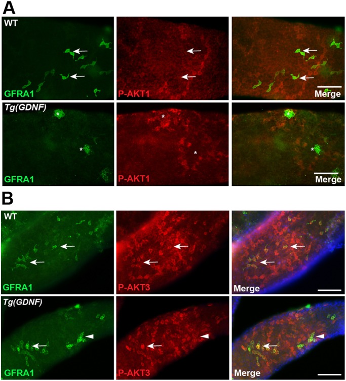
AKT1 and AKT3 are phosphorylated in different types of spermatogonia. (A) Whole-mount wild-type or Tg(Gdnf) tubules co-immunostained for phosphorylated AKT1 Ser473 (P-AKT1) and GFRA1. GFRA+ cells in wild-type testes (arrows) and large clusters of GFRA+ cells in Tg(GDNF) testes (asterisks) are P-AKT1 negative. (B) Whole-mount wild-type or Tg(Gdnf) tubules co-immunostained for phosphorylated AKT3 Ser472 (P-AKT3) and GFRA1. Arrows indicate GFRA1+ P-AKT3+ As and Apr spermatogonia. Arrowheads indicate GFRA1+ P-AKT3+ cluster in Tg(Gdnf) tubules. See also Figs S5 and S6. Scale bars: 100 µm.
