Fig. 1.
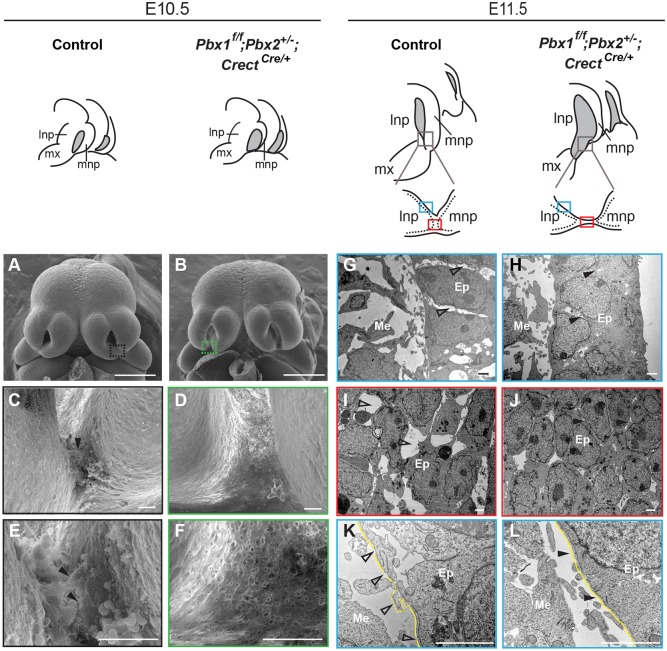
Epithelial morphological changes at the lambdoidal junction (λ) are Pbx dependent. Top: sketches of embryonic midfaces of indicated genotypes. Nasal pits are shown in gray. Plane of sections of control and mutant λ are shown below the E11.5 sketches. Dotted lines indicate epithelium; blue rectangles indicate the sectioned region through lnp at the λ (G,H,K,L); red rectangles the sectioned region through lnp/mnp fusion (I,J). (A-F) SEM of control (A) and mutant (B) heads. λ domains (black and green rectangles) are shown at higher magnification in C,E (control) and D,F (mutant). Filled black arrowheads indicate cellular protrusions at the lnp/mnp fusion site (C,E). (G-L) TEM of control (G,I,K) and mutant (H,J,L) λ sections through planes in blue and red rectangles, respectively. (G-J) Empty space enlargements between epithelial cells are shown by unfilled arrowheads in control (G,I). Close contacts between epithelial cells are indicated by filled arrowheads in mutant (H,J). (K,L) In control (K), note break down (unfilled arrowheads) and blebs (dotted line) of BM (yellow line) whereas in the mutant (L) BM is intact (arrowheads). Scale bars: 500 µm (A,B); 20 µm (C-F); 2 µm (G-L). Ep, epithelium; Me, mesenchyme; mx, maxillary process.

