Fig. 7.
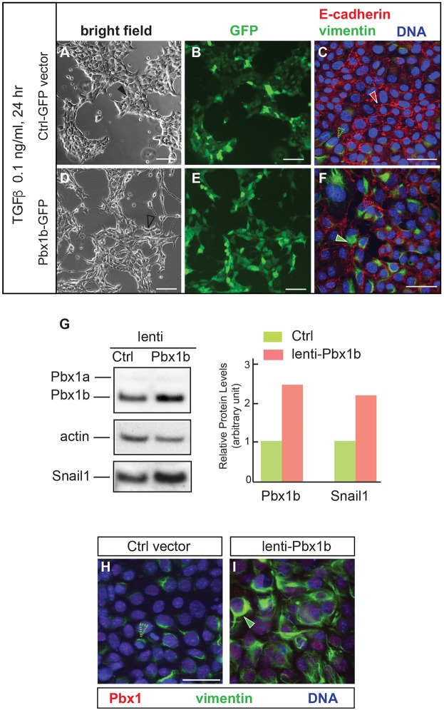
Forced Pbx1 expression is sufficient to induce EMT in NMuMG cells. (A-F) Morphological alterations and changes in EMT signatures in TGFβ-treated cells (0.1 ng/ml for 24 h) overexpressing Pbx1b, versus control. (A,D) Bright field shows shape changes and scattering in NMuMG cells transduced with a lentivirus carrying mPbx1b (unfilled black arrowhead) versus control (Ctrl; filled black arrowhead). (B,E) GFP visualizes similar transfection efficiency in live cells with or without mPbx1b overexpression. (C,F) Immunofluorescence shows membrane-associated E-cadherin (filled red arrowhead) and negligible filamentous vimentin (unfilled green arrowhead) in fixed cells transduced with control vector (C). Increase of vimentin (filled green arrowhead) and E-cadherin reduction at cell junctions (unfilled red arrowhead) indicate EMT induction in mPbx1b-lentivirus transduced cells (F). (G) Western blot of Pbx1a, Pbx1b and Snail1 levels in NMuMG cells transduced with Ctrl or mPbx1b. Pbx1a is present at low levels. Loading control: actin. Quantification of western blot is shown on the right. (H,I) Immunofluorescence of fixed cells transduced with lentivirus carrying Ctrl (H) or mPbx1b (I) construct for Pbx1 (red) and vimentin (green). DAPI (blue) stains nuclei. In controls, a small number of cells with low endogenous vimentin were observed (unfilled green arrowhead, H). In cells overexpressing mPbx1b, induction of filamentous cytoplasmic vimentin was observed (green arrowhead, I). Scale bars: 50 µm (A,B,D,E); 25 µm (C,F,H,I).

