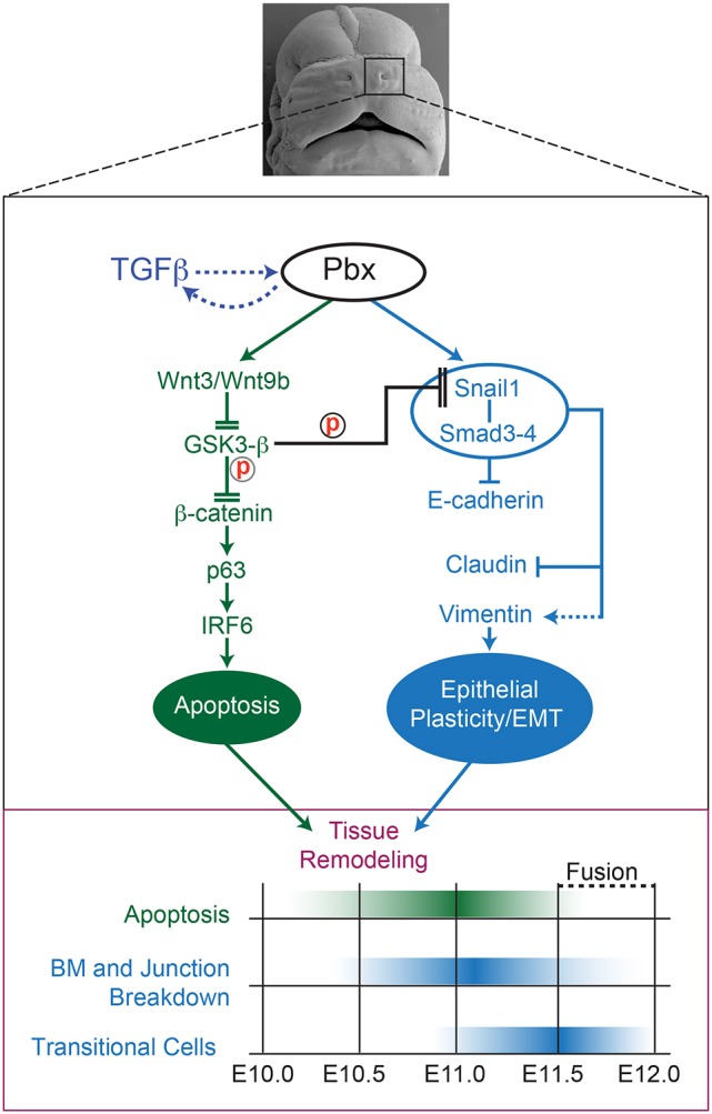
Fig. 8.

Pbx-dependent crosstalking regulatory axes direct dynamic cellular behaviors controlling epithelial tissue remodeling at the λ. Top: Frontal view of E13.5 murine midface after lnp/mnp/mx fusion. Black rectangle (enlarged in middle panel) highlights Pbx-dependent regulatory networks (active from E10.0 to E12.0). Cross-regulatory loop between TGFβ and Pbx is indicated by dashed arrows. In NMuMG cells, TGFβ promotes Pbx nuclear accumulation (see Fig. 5) and, concomitantly, in the embryo Pbx1/2 loss of function results in loss of Smad4 at the λ epithelia (see Fig. 3). The green axis controls epithelial apoptosis (Ferretti et al., 2011), whereas the blue axis mediates epithelial plasticity/EMT at the λ. These regulatory axes can crosstalk via Snail1 post-transcriptional control by GSK3-β (Zhou et al., 2004; Yook et al., 2006) and converge to execute tissue remodeling. Solid arrows indicate direct transcriptional control; dashed arrows indirect activation; flat heads transcriptional repression; double flat heads reduced protein activity through post-transcriptional mechanisms. Bottom: Developmental timing of epithelial tissue remodeling. Green hue highlights the peak of apoptosis; blue hue the breakdown of the BM and adherens and tight junctions, as well as the appearance of transitional cells. ‘Fusion’ indicates the replacement of the frontonasal epithelial seam by the mesenchymal bridge from E11.5 to E12.0.
