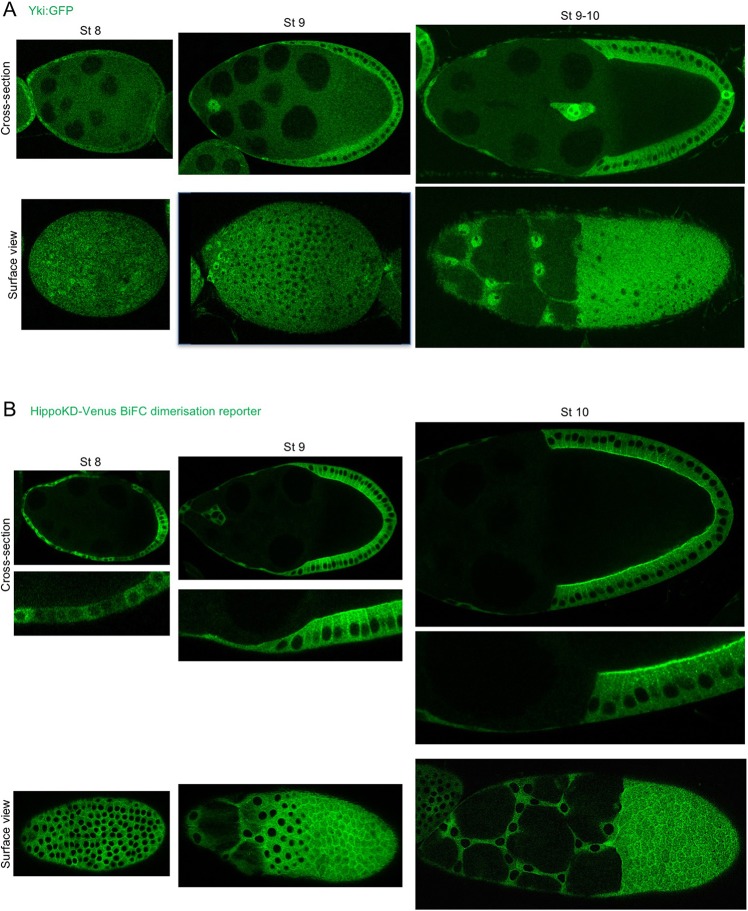
Fig. 3.
Yki translocates to the nucleus in stretch cells and to the cytoplasm of columnar cells, inversely correlating with Hippo dimerisation at the apical plasma membrane. (A) Endogenous Yki with a GFP tag was visualised in the views shown at different stages of oogenesis. At stage 8, Yki:GFP is predominantly cytoplasmic in the cuboidal epithelium, but at the anterior where the cells start to flatten Yki:GFP is found in the nucleus. During stage 9, Yki:GFP can clearly be found in the nucleus of stretch cells and in the cytoplasm of columnar cells. This pattern is even more pronounced at stage 10. (B) The HippoKD-Venus dimerisation reporter was visualised in the views shown over different stages of oogenesis. At stage 8, a weak Hippo dimerisation signal is observed with an apical signal apparent in the posterior epithelium. At stages 9 and 10, a clear apical and lateral signal can be seen in columnar cells, whereas in stretch cells there is a faint cytoplasmic signal.

