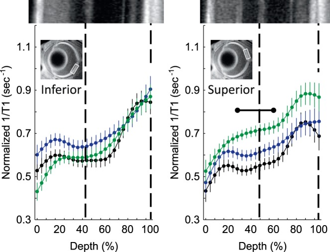
Figure 5.

Excessive free radical production in female dark-reared P30 mice retina. Modeling results of normalized 1/T1 MRI profiles in vivo for central retina on either the inferior (left) or superior (right) side (indicated by the box in the MRI insert of each graph; see Methods for details) for the following groups: dark-reared P30 wt group (black, n = 6), dark-reared P30 Pde6brd10 mice injected with saline (green, n = 6), and dark-reared P30 Pde6brd10 mice injected with MB (blue, n = 5). Other figure conventions are detailed in Figure 1.
