Figure 5.
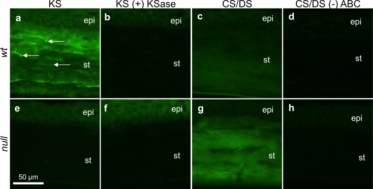
Immunofluorescent labeling of low-sulfated KS- and CS/DS-GAGs in WT (a–d) and null (e–h) corneal tissue. WT tissue labeled for KS-GAGs fluoresced at all stromal depths and featured occasional bright punctae (white arrows, a); this signal was sensitive to keratanase (KSase) digestion (b). The lack of fluorescent signal in the null corneal stroma, without (e) or with (f) KSase incubation, implied a lack of sulfated KS-GAGs in the mutated corneal stroma. Chondroitinase ABC-liberated CS/DS-GAG “stubs” were local to WT (c) and null (g) tissue throughout the depth of the corneal stroma. A brighter stromal fluorescent signal from the null specimen, relative to the WT control, indicated increased CS/DS content in the B3gnt7 knockout. In absence of chondroitinase ABC predigestion, neither WT (d) nor null (h) tissue appeared positive for the 2B6 stub epitope. Epi, corneal epithelium; st, stroma. All images were collected at 40× magnification. Scale bar denotes 50 μm.

