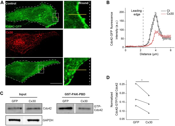
Fig. 4.
Cx30 controls the recruitment and activation of Cdc42 in migrating astrocytes. (A) Primary astrocyte cultures were transfected with Cx30 and GFP-Cdc42. 8 h after wounding, cells were immunostained for Cx30 and the intensity of the GFP-Cdc42 signal at the leading edge was measured. Scale bar: 20 µm. (B) Quantitative analysis of the linear intensity profile of GFP-Cdc42 at the leading edge. Cx30 reduced significantly the recruitment of GFP-Cdc42 at the leading edge (Ct, n=19 cells; Cx30, n=15 cells). (C) Cdc42 pull-down activation assay in astrocytes transfected with Cx30 or GFP 30 min after wounding. Immunoblots showing Cdc42 protein levels in the total (Input) and GTP-bound fractions (GST-PAK-PBD). GAPDH (total) and Ponceau staining (GTP) were used as loading controls. (D) Quantitative analysis of Cdc42 activation (n=3 cultures). Cdc42-GTP values were normalized to Cdc42 levels in the total fractions. Cx30 inhibited Cdc42 activation. Asterisk indicates statistical significance (*P<0.05, paired t-test).

