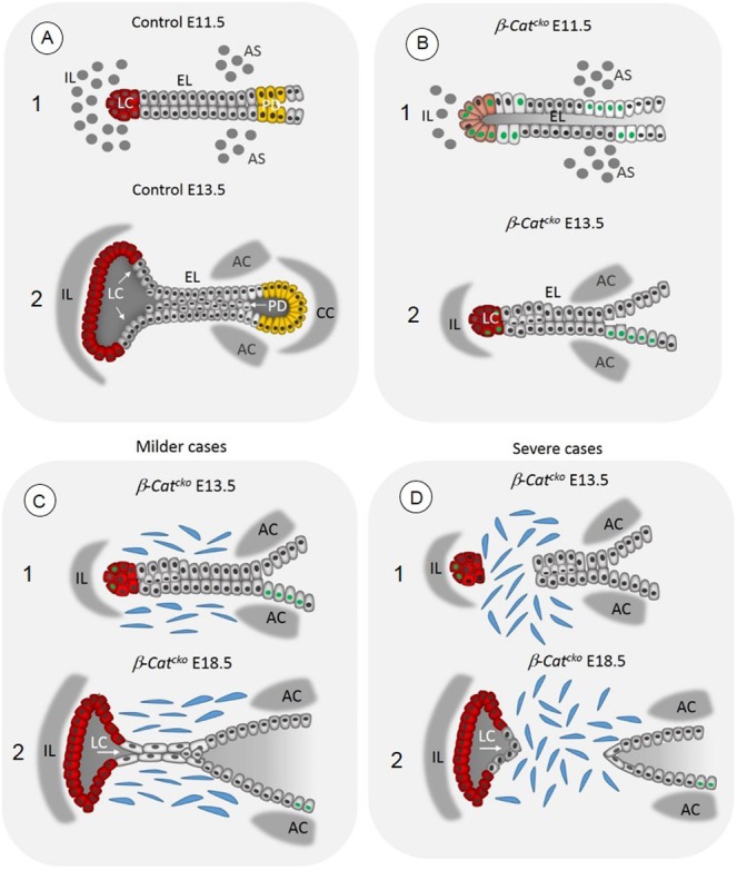
Fig. 8.

Schematic illustration demonstrating the EL fusion and recanalization in control embryos and β-Catcko mutants, in milder and more severe cases of laryngeal webbing. (A1) Formation of the EL in control embryos at E11.5. (A2) Formation of LC (red cells) and PD (yellow cells) in control embryos at E13.5. Epithelial cells in the EL express p63 (gray nuclei), proliferate and accumulate along the EL midline. Meanwhile mesenchymal cells differentiate into laryngeal cartilages. (B1) Inactivation of β-catenin in β-Catcko mutants leads to the failure in the EL fusion at E11.5 and disrupted initial differentiation of VF basal progenitors. Some VF epithelial cells remain columnar and fail to upregulate p63 (green nuclei). (B2) The delay in the EL fusion leads to the delay in LC expansion (in red) and the absence of the PD and the septum with the cricoid cartilage. (C) Failure in specification of basally positioned of VF progenitors versus a suprabasal cell layer may prevent EL recanalization in milder web cases. (D) In more severe cases of laryngeal webs, inactivation of β-catenin in the epithelium affects the EL integrity. After delayed EL fusion, cells at the tip of the EL can pinch off from the remaining EL, enabling mesenchymal cells to migrate into the space in between. It is also possible that some epithelial cells undergo aberrant epithelial-mesenchymal transition and contribute to laryngeal web formation. AS, arytenoid swelling; AC, arytenoid cartilage; CC, cricoid cartilage; EL, epithelial lamina; IL, intermediate lamina; LC, laryngeal cecum; PD, pharyngoglottic duct.
