. 2018 Mar 12;7:e26016. doi: 10.7554/eLife.26016

Figure 6. Connectome of DnB to premotor and nociceptive interneuron outputs.

(A) First instar larval CNS showing reconstruction of DnB neurons (green), and nociceptive integrating interneurons (purple). Output synapses are indicated in red and input synapses in cyan. Nociceptive interneurons primarily receive input from output sites on DnB dendrites. (B) Percent of top hits’ (>3 synapses) output from right (DnB a1R) and left (DnB a1L) A1 DnB neurons as a function of cell type. Premotor circuits and nociceptive integrators are dominant outputs of DnB neurons. (C) Identities of nociceptive integrating targets for right and left DnB neurons in A1. Numbers of synapses reconstructed are indicated. Width of arrow corresponds to degree of synaptic connectivity. (D) First instar larval CNS showing reconstruction of DnB neurons (green), premotor (purple), and motor targets (yellow). Premotor neuron output synapses (red dots) located primarily in motor domain (arrowhead). (E) Identities of premotor targets for right and left DnB neurons in A1. Numbers of synapses reconstructed are indicated. Dominant outputs are A27k and A01d-3 premotor neurons. Width of arrow corresponds to degree of synaptic connectivity. (See also Figure 6—figure supplement 1).
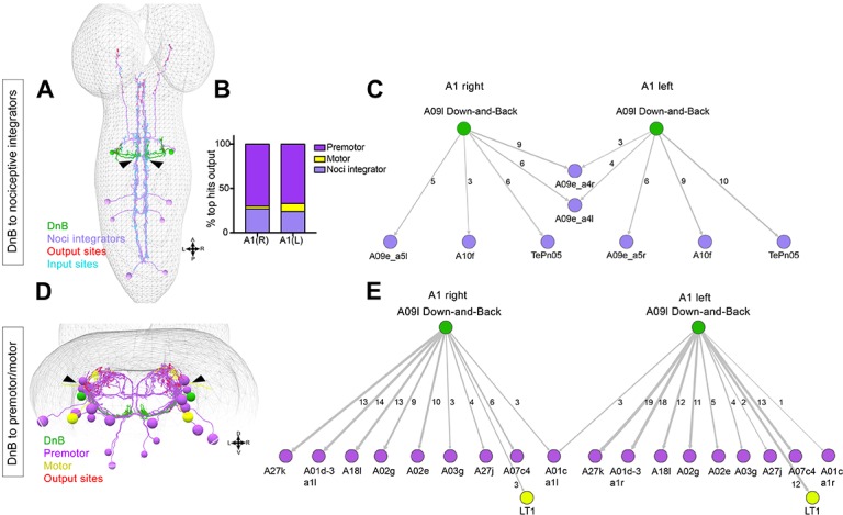
Figure 6—source data 1. Summary table for output connectivity graph.Percentage of top hit neurons (>3 synapses with DnB) that fall into the category: premotor, motor, or nociceptive integrator neurons.
DOI: 10.7554/eLife.26016.026

Figure 6.

Figure 6—figure supplement 1. Additional properties of Down-and-Back output signaling.
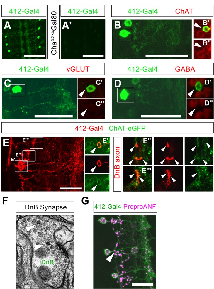
Figure 6—figure supplement 1.

(A) mCD8:GFP driven by 412-Gal4, labeled with anti-GFP (green). (A') Cha3.3kb-Gal80 reduces expression of mCD8:GFP (anti-GFP, green) in DnB neurons. (B) DnB neuron (anti-GFP, green); (C') Boxed region: DnB cell body (arrowhead) shows overlap with ChAT (anti-ChAT; dsRed), (C'') ChAT; dsRed channel only. (C) DnB neuron (anti-GFP, green); (B') Boxed region: DnB cell body (arrowhead) shows no overlap with vGLUT (anti-vGLUT; dsRed); (B'') VGLUT; dsRed channel only. (D) DnB neuron (anti-GFP, green); (D') Boxed region: DnB cell body (arrowhead) shows no overlap with GABA (anti-GABA; dsRed), (D'') GABA; dsRed channel only. (E) tdTomato driven by 412-Gal4 labeling DnB neuron (anti-dsRed, red) (D') Single section showing DnB cell body overlapping with ChAT-eGFP (anti-GFP, green) (top: overlap, middle: tdTomato, red channel only, bottom: ChAT-eGFP cell body) (D''–D''') Single sections showing DnB axons; left panels DnB axon and ChAT-eGFP puncta overlap; middle: DnB axons, tdTomato channel; right panels: ChAT-eGFP puncta, GFP channel only. (F) Electron micrograph of DnB synapse showing dense core vesicles (arrowheads) indicative of peptidergic or aminergic release. (G) Neuropeptide precursor, PreproANF fused with emerald GFP (anti-GFP, magenta) is processed and expressed in DnB axons (arrowhead; anti-dsRed, green), suggesting that DnBs are peptidergic. Genotypes: (A,B,C) 412-Gal4, UAS-mCD8:GFP /+ (A') 412-Gal4, UAS-mCD8:GFP/Cha3.3Kb-Gal80 (D–D''') 412-Gal4, UAS-TdTomato/Chat-EGFP-Flash-Tev3xFlag. (G) UAS-Prepro:ANF-EMD;;412-Gal4,UAS-TdTomato/+. Scale bars = 50 µm (A– A') 20 µm (B,C,D,G).