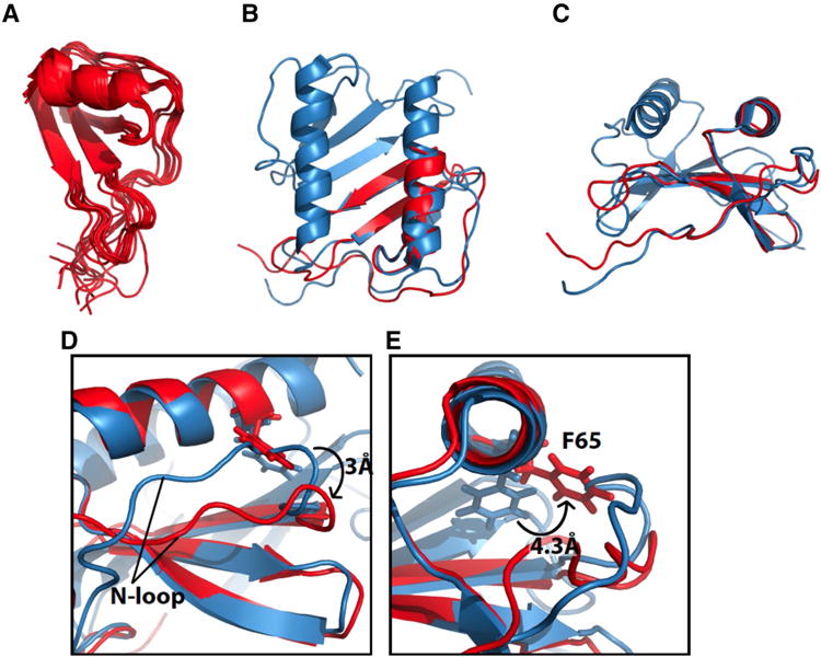
Fig. 3.

Solution NMR structure of monomeric IL-8 (1–66). a Overlay of the ten lowest energy structures of monomeric IL-8(1–66). b Structure of monomeric IL-8(1–66) in red overlayed with the solution NMR structure of full-length IL-8(1–72) in blue (PDB ID: 1IL8). c Same as in b only rotated 90°. d Expanded structural region of monomeric IL-8(1–66) in red and wild type IL-8 in blue. The change in position of the N-loop relative to the C-terminal helix between monomeric IL-8 and wild type IL-8 is shown. Differences in distances as measured between the amide nitrogen atoms of residues K20 and F65 e Expanded structural region of monomeric IL-8(1–66) in red and wild-type IL-8 in blue. The difference in position of the C5 atom of Phe65 between monomeric IL-8(1–66) and wild type IL-8(1–72) is shown
