
Figure 2.
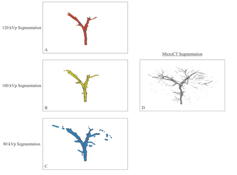
Segmentation of imaging volumes with CBCT at 120 kVp (A), 100 kVp (B) and 80 kVp (C) and microCT (D). The microCT segmentation shows more extensive ROB distribution than the CBCT segmentations. Among the CBCT segmentations, visualization of distal vasculature improved as kVp decreased.