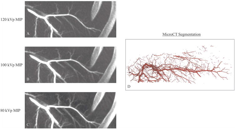
Figure 4.

Comparison of MIPs generated from CBCT images acquired at 120 kVp (A), 100 kVp (B) and 80 kVp (C) with the corresponding microCT segmentation (D). Presented MIPs were cropped from the full in vivo image to highlight the area corresponding to the explanted sample. Improved visualization of distal vasculature was noted in MIP images as CBCT kVp decreased.
