Figure 5.

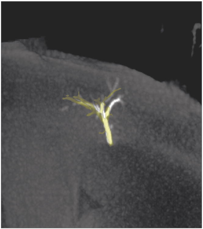
100kVp CBCT (yellow) segmentation and 40 mm slab MIP. Improved visualization of distal vasculature was observed on the MIP versus the CBCT segmentation.

100kVp CBCT (yellow) segmentation and 40 mm slab MIP. Improved visualization of distal vasculature was observed on the MIP versus the CBCT segmentation.