Figure 4.

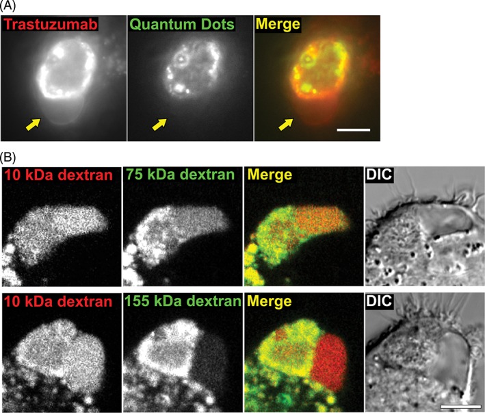
The vacuole is separated from the phagosome by a semi‐permeable barrier. A, MDA‐MB‐453 cells were opsonized with Alexa 488‐labeled trastuzumab (pseudocolored red) and counterstained with anti‐human Fab‐conjugated QDot 655 quantum dot nanoparticles (pseudocolored green). The cells were co‐incubated with J774A.1 macrophages for 6 hours, followed by imaging (fluorescence). Yellow arrows indicate the location of the vacuole. B, MDA‐MB‐453 cells were opsonized with trastuzumab and co‐incubated with J774A.1 macrophages for 6 hours followed by imaging (fluorescence and DIC). The macrophages were preloaded with Alexa 647‐labeled 10 kDa dextran (pseudocolored red) and TRITC‐labeled dextran of 75 or 155 kDa molecular weight (pseudocolored green) as indicated. Images of representative cells from at least 22 cells and 2 independent experiments are shown. Scale bars = 5 μm
