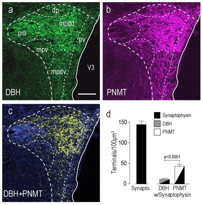
Figure 6. Catecholaminergic Innervation to the PVH.
DBH (a) and PNMT (b) immunoreactivity in the rat PVH and its surrounding regions. Panel c) is a merge of a) and b) to show co-occurring DBH and PNMT structures and DBH-only structures. Within the PVHmpdd there are significantly more PNMT than DBH- only structures (c). Scale bar is 150μm for all panels. Abbreviations are as Figure 1 except for: mpdd, medial parvicellular part, dorsal zone (dorsal division); mpdv, medial parvicellular part, dorsal zone (ventral division). Panel d) shows that there were significantly more PMNT-containing presynaptic terminals in the PVHmpd than those containing DBH-only (no confocal images are shown for these results). Error bars indicate mean + S.E.M.

