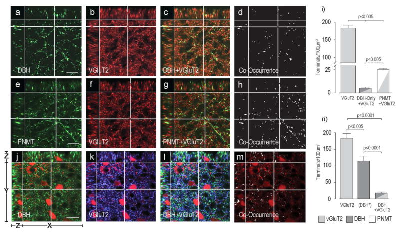
Figure 7. Some Catecholamine Terminals in the PVHmpd of the Rat and Mouse Express VGluT2.
Panels a) – h) show DBH-ir (a–d) or PNMT-ir (e–h) in the rat PVHmpd together with VGluT2-ir (b,f) and its co-occurrence with DBH (c,d) or PNMT (g,h). There are significantly more PNMT than DBH-only terminals that also contain VGluT2 in the rat PVHmpd (i). Please note the change in the scale of the Y axis in panel i). Panels j) – m) show DBH-ir (j) and VGluT2-ir (k) in the female mouse PVHmpd. TdTomato-labeled CRH soma can be seen in panels j) – m). The co-occurrence of DBH and VGluT2 is shown in l) and m). Scale bar indicates 20μm for all panels. Note that in panel n) the (DBH*) data are DBH-immunoreactive structures only. Group data in n) are from female mice. Because of species incompatibility there was no synaptophysin labeling in this experiment to verify that non-VGluT2-ir DBH structures were terminals. Scale bar indicates 20μm for all image panels. Please see Figure 2 for more information about the general organization of the confocal image panels. Error bars indicate mean + S.E.M.

