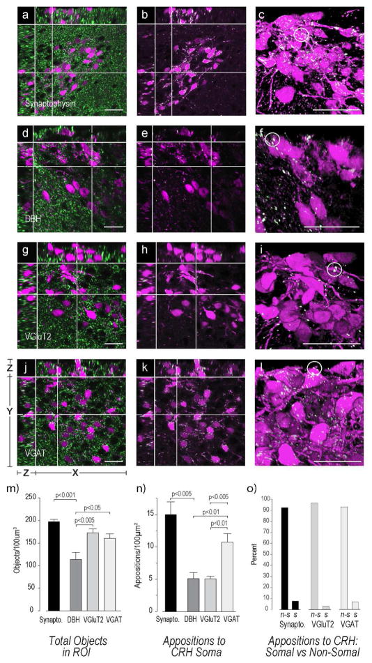
Figure 9. Appositions to CRH Neurons in the Mouse PVHmpd.
Female mouse sections containing the PVHmpd were labeled with antibodies against synaptophysin (a–c), DBH (d–f), VGluT2 (g–i), and VGAT (j–l) and analyzed for appositions of each type against the soma of the CRH neuron. Please see Figure 2 for more information about the general organization of the confocal image panels. The white structures in all confocal images except a) d), g), and j) are those that are double-labeled and fulfill the apposition criterion established by Bouyer & Simerly [2013]. See Methods for more details. Images c), f), i), and l) are 3D renderings of the Z-stacks used for images b), e), h), and k), respectively. White circles highlight some somatic appositions. There are significantly more VGAT and VGluT2 terminals than DBH terminals (m). However, VGAT somatic appositions are more numerous than either VGluT2 or DBH (n). There are significantly more appositions to the non-somatic regions of the CRH neuron (o). Group data in m) – n) are from female mice. Abbreviations for o): n-s, non-somatic; s, somatic. Scale bars indicate 20μm. Error bars indicate mean + S.E.M.

